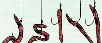
Reproduction of earthworms
Well-known earthworms are not only one of the links in the food chain in nature. These are also mini-factories for processing organic residues that fall on the ground into a nutritious substance called humus. Thanks to them, the soil in which they live becomes richer and is able to breathe. The underground passages that the worm makes as it moves below the surface allow air to penetrate to the roots of the plants.
The meaning of flatworms
Ciliated worms provide food for many animals. Many types of flatworms are parasites and cause severe diseases in animals and humans. Diseases caused by parasitic worms (helminths) are called helminthiasis . The severity of the disease most often depends on the number of parasites that have entered the body. Helminths can live in almost all organs. It is necessary to carry out preventive procedures to prevent the spread of helminthiases.
Head-tailed or tail-headed?
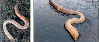
If you don’t know the body structure of an earthworm, you can very easily make a mistake when determining where its head is and where its tail is. Its elongated shape without any external organs allows the worm to calmly move through the earth in search of food.
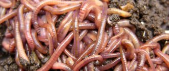
Earthworms are a type of annelid worms, the very name of which indicates the characteristic structure of their body. They seem to be assembled from small rings strung on an elastic band. The segments formed by the constrictions have small bristles on the sides, with the help of which the worm moves.
Unlike many others, the earthworm does not have many such setae, for which its subclass received the name oligochaete. In each individual, closer to the head part, the body is surrounded by a thickening, which gave the name to the class - girdle . Its presence indicates the worm's ability to reproduce. This belt is part of the reproductive system of these animals.
Alternative ways to survive
If a situation suddenly arises when a species is threatened with complete extermination, earthworms have backup options that help them survive in the most difficult conditions. These animals are able to reproduce without fertilization. But then there will be only one females in the population.
There are species that can reproduce asexually. If you cut such a worm, then each half will grow back the missing part.
To understand how worms reproduce, you need to have some idea of the environment in which they live and their structure.
Under natural conditions, raincoats colonized the upper soil layer. These animals have no division into males and females; they are hermaphrodites.
Hermaphrodite is not a dirty word
All adult earthworms simultaneously carry the characteristics of the sex cells and glands of the male and female. The scientific name of such an animal is hermaphrodite . Representatives of such species can reproduce if there are only two living organisms. It doesn't matter to them what gender these organisms are. Thanks to this property, this species is distributed everywhere and is very numerous, moreover, it has many related species.
Reproduction of earthworms occurs in several stages. First, individuals exchange seminal fluid, which will be stored for a certain number of days for each species in mucus secreted by special cells of the girdle. When the sperm mature, the same girdle again secretes mucus, which forms a cocoon. The worm removes it from itself through the head, and during the movement, eggs enter the cocoon from the genital segments. And only now their fertilization .
The removed cocoon, filled with viable eggs, is stored in the ground, where it develops new earthworms. When the right time comes, small but already fully formed worms emerge from such a capsule.
General characteristics of flatworms
Subkingdom multicellular. Type of flatworms. Features of life activity: structure, integument, nervous system, sensory organs, nutrition, respiration, excretion, reproduction and diversity (class Ciliated worms, class Flukes and class Tapeworms)
Over 12 thousand species are known that belong to the type Flatworms (Plathelmintes or Platodes). They have bilateral (bilateral) symmetry (appears for the first time in multicellular organisms). They live mainly in seas and fresh water bodies. There are parasitic forms.
The body is flattened in the dorsal-ventral direction. There are anterior and posterior ends. Flatworms and representatives of all other types are three-layered animals. In addition to the ecto- and endoderm, in the process of individual development, an intermediate, or third germ layer, the mesoderm, is formed.
The integument of the body forms a skin-muscular sac of epithelium and muscle tissue (consists of several layers of muscle fibers that are not collected into separate muscle bundles). The body consists of 3 layers (external, middle, internal). Muscles are smooth (3 layers: circular, oblique, longitudinal). They have no body cavities. These are parenchymatous animals. The spaces between the internal organs are filled with loose connective tissue - parenchyma, which performs various functions: supporting, excretory, transport of substances, storage of nutrients, excretory function.
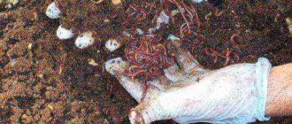
Breeding and propagation of earthworms at home.
The author of the work was awarded a third degree winner diploma
I. Management
One of the many Chinese wisdoms says that it is not the farmer who grows a good harvest who is rich, but the one who cultivated the land that gave it. In the last century, the lands of Russia were depleted by the intensive use of mineral fertilizers, and today organic farming has only just begun to revive. It is no secret that soil rich in organic matter of plant and animal origin is the most fertile, and ordinary earthworms make it so. They are the ones who process the remains of organic compounds into valuable fertile soil called humus.
Earthworms in the soil are a factor in its fertility and a condition for the normal development of plants. Earthworms - process, fertilize and increase productivity, not only consuming from their habitat, but giving tens of times more, helping industry and agriculture, controlling the biological balance of nature
Many people do not know the priceless virtues of these workers of the earth, and sometimes consider earthworms to be harmful creatures. This misconception arose from ignorance.
Variety of flatworms
Parasitic flatworms: Pork tapeworm and Liver fluke
This type includes 5 classes, four of which are exclusively parasitic forms.
Class Ciliated worms (Turbellaria)
Type of flatworms. Planarian structure: external structure, digestive, nervous, reproductive and excretory systems. Planaria capturing prey
About 3 thousand species are known. The majority are free-living. Distributed in seas, fresh water bodies, sometimes in moist soil or on its surface. The body is covered with cilia. Dimensions range from 1 mm to several centimeters. Most animals have bright colors due to the presence of pigment in the skin. Small animals move with the help of cilia and muscle contractions, large animals move only with the help of muscle contractions. The cavities between the organs are filled with loose parenchyma. Between its cells there is a watery fluid, which allows the parenchyma to act as an intermediary between the intestines and internal organs. The digestive system consists of the foregut and middle intestine, which are blindly closed. Predators feed mainly on invertebrates. Only a few have adapted to a parasitic lifestyle (ectoparasites of crustaceans, mollusks and turtles). The nervous system consists of a double node - the cerebral ganglion and the nerve trunks extending from it. Sense organs are developed: balance organs ( statocysts ), light-sensitive eyes (one or several pairs), tactile cells.
Representatives: white or milky planaria, black multi-eyed planaria , etc.
White or milky planaria
Planaria. Photo of planaria. The planaria captured the crustacean. Digestive, nervous, reproductive and excretory systems of planarians. Cross section of planaria and reproduction of planaria by division
Lives in fresh water bodies with stagnant water. It has a milky white color. Dimensions: approximately 25 mm long, 6 mm wide.
The body is leaf-shaped, covered with cilia. The head end of the body has two lateral lobes, at the base of which there is a sucking pit - a fixation organ. The musculocutaneous sac consists of single-layer epithelium and longitudinal, circular and dorso-abdominal muscles. In the cytoplasm of epithelial cells there are special rod-shaped structures ( rhabdoids ). If the animal is disturbed, they are thrown out of the cage, break down and form a protective layer of mucus on the surface. Among the epithelial cells at the edges of the body there are unicellular glands or gland ducts, the body of which is immersed in the parenchyma. There is no body cavity. The spaces between the internal organs are filled with parenchyma.
The mouth opening is on the ventral side in the middle of the body. The pharynx looks like a proboscis. The middle section of the intestine is represented by three branches, one of which is directed forward and two directed backward. All of them produce a large number of side branches. Blindly closed. Digestion – intracellular and extracellular. The white planaria is a predator. Feeds on small aquatic animals. Undigested food remains accumulate in the intestines and are then thrown out through the mouth.
Respiratory organs and circulatory systems are absent. Breathing through the entire surface of the body. The excretory system is represented by two branched tubules located on the sides, into which excretory products come from protonephridia.
The nervous system is formed by the head nerve ganglion from several nerve trunks, of which two lateral, connected circular nerve jumpers are well developed. Sense organs - light-sensitive eyes, tactile cells (sensilla), two tentacles on the front of the body. Planaria have two eyes. The balance organ is located between the eyes in front of the brain.
Hermaphrodite. The genital opening is located on the abdominal part of the body. Fertilization is cross-fertilization. After fertilization, it lays eggs in a cocoon, where the offspring develop. Development is direct. Has good regeneration ability.
Class Flukes or Trematodes (Trematoda)
Over 4 thousand species are known, over 600 of which are in our country. Parasites (endo- and ectoparasites) that live in or on the body of humans and animals. Parasitic worms, regardless of classification, are called helminths . They don't have eyelashes. Many sucking species have small spines that cover the entire body and facilitate attachment to the host. They are characterized by the presence of two suckers: the oral one, at the bottom of which there is a mouth opening, and the abdominal one. They have a leaf-shaped form. The body is covered with a shell of compacted cytoplasm of epithelial cells, which performs a protective function. Underneath it are layers of circular, oblique and longitudinal muscles. The digestive, nervous and excretory systems have a structure typical of flatworms. Sense organs are poorly developed. The reproductive system is well developed. Mostly bisexual (hermaphrodites). They have high fertility. The life cycle is accompanied by a change of hosts (1 – 2 intermediate and the main one).
Representatives: liver fluke, Siberian or cat fluke, pulmonary fluke , etc.
Liver fluke (Fasciola hepatica)
The structure and development cycle of the liver fluke
It has a yellow-green color with reddish-brown edges. Body length is 3-5 cm.
The head end is narrowed. The oral sucker is located here. On the ventral side of the body lies an additional ventral sucker, which is an organ of attachment and has no connection with the digestive system. Between the suckers there is a mouth opening, and next to it there is an excretory pore. The body is not dissected, leaf-shaped. There is no body cavity.
The digestive system includes the anterior (oral opening, pharynx, esophagus) and middle (two parallel lateral canals, which are strongly branched and blindly closed) intestines. Undigested remains are expelled through the mouth. The nervous system consists of the peripharyngeal ganglia and three pairs of nerve trunks extending from them. The most developed is one pair, which stretches along the sides of the body along the ventral side.
Hermaphrodite. Has a complex reproductive system. The uterus is small, rosette-shaped and located behind the abdominal sucker. The testes and ovaries are located behind the uterus, very branched. The eggs are yellow-brown in color. It can produce over 100 thousand eggs per day.
It parasitizes the liver and bile ducts of herbivores (cattle, sheep, pigs, goats, horses), and sometimes humans. The intermediate host is the small pond snail. Infection occurs when drinking water from reservoirs. The parasite eggs are hatched from the host's intestines. They need to get into the water. miracidia emerge from them , covered with cilia, freely swimming, which then penetrate the body of the small pond snail. The larva burrows into the lung tissue with the help of a proboscis, and then settles in the liver. In the body of the mollusk, the larva loses its cilia, develops into a new larval stage ( sporocyst ), reproduces parthenogenetically (without fertilization) and forms a large number of new larvae ( redia ). Such larvae have a mouth opening, a pharynx and an intestine. They develop special rudimentary cells, from which, under favorable conditions (warm seasons), another larval stage ( cercariae ) is formed. These larvae emerge from the body of the mollusk and have muscular tails, with the help of which they swim to the shore, attach to plants, or stay at the surface of the water. The tail is destroyed. The body of the larvae is covered with a shell. They become motionless. They enter the intestines of animals with water and grass. Here the larvae are freed from the shell, drill through the intestinal wall and enter the abdominal cavity, from where they migrate to the liver. They penetrate through the liver tissue into the bile ducts, where after 3 to 4 months they become sexually mature and begin to produce eggs. The parasite leads to degeneration of the bile ducts, destruction of liver tissue and inflammatory processes. Sometimes parasites can block the bile ducts. The body is poisoned by the fluke's metabolic products. The disease is accompanied by fever, severe pain in the liver, etc. A person can become infected by drinking raw water, eating sorrel, watercress, etc.
Cat fluke (Opisthorchis felineus)
It parasitizes the liver and bile ducts, sometimes in the pancreatic ducts of animals (dogs, cats, etc.) and humans. Length – 8 – 13 mm. The two testes are located at the back of the body and are rosette-shaped. The life cycle occurs with the help of two hosts: betinia (a freshwater gastropod) and freshwater cyprinid fish.
To prevent the disease, it is necessary to treat patients and eat well-processed fish.
Class Tapeworms or Cestodes (Cestoda)
Class Tapeworms. General view of a tapeworm. Pork tapeworm, ox tapeworm and broad tapeworm
About 3,500 species are known, 500 of which are known in our country. All species are exclusively parasites. The shape of the body resembles a ribbon, for which they received the corresponding name. Sizes range from 1 mm to 10 meters or more. They parasitize in the sexually mature state in the intestines of humans and vertebrates, and in the larval state - in the tissues and organs of invertebrates or vertebrates.
The body ( strobilus ) consists of a head ( scolex ), on which the organs of attachment are located, an undivided portion - the neck, and a dissected part - the segments. Possible organs of attachment are suction cups, suction slits, hooks. The segments are formed at the posterior end of the neck. The number of segments ranges from 3 - 4 (in Echinococcus) to 40 thousand or more (in the broad tapeworm).
They do not have sensory organs, circulatory, respiratory or digestive systems. Nutrients are absorbed from the intestines by the entire surface of the body. Digestion is intracellular. Parasites are surrounded by semi-digested food of their hosts, so the body is covered with microscopic growths that increase its absorption surface.
Isolation is carried out with the help of protonephridia. The main excretory canals (two on each side) run along the edges of all segments from the head to the end of the strobila. In the parenchyma there are limestone cells with accumulated waste products.
An independent hermaphroditic reproductive apparatus develops in each segment. The youngest segments are near the neck, the oldest are at the posterior end, where they come off. There is no reproductive system in the segments that are located closer to the neck. First, the male genital organs are formed, then the female ones (the segment becomes hermaphroditic). Mature eggs are located in the last segments, which can either be thrown out passively with feces, or crawl out on their own. The uterus has no outlet. The last segments of the body with eggs and feces enter the environment. The eggs (segments) enter the stomach of the intermediate host (cattle, pigs, humans, etc.). From the eggs hatch larvae with hooks ( oncosphere ), with the help of which they enter the blood through the intestinal wall and are carried into the muscles and organs, where they turn into a fin (a vesicle in which the head of the worm is located). The main host becomes infected when it eats raw, poorly fried, or cooked meat or fish. The symptoms of diseases that cause cestodes are similar: insomnia, weakness, nausea, seizures similar to epileptic, etc.
Representatives: bovine tapeworm, pork tapeworm, broad tapeworm and others.
Bull or naked tapeworm (Taeniarhynchus saginatus)
The structure and development cycle of the bovine tapeworm
Has a whitish color. The body is ribbon-shaped. Length – up to 10 – 12 m. There are four suckers on the head. Segments ( proglotids ) - about a thousand. The muscles are quite strong, which is associated with significant mobility of the parasite in the intestines. The adult worm lives in the human small intestine (the definitive host). The intermediate host (the host in whose body the larval stages develop) is cattle.
The digestive, circulatory systems, sensory organs, and breathing are absent. Nutrients are absorbed by the entire surface of the body. The integument is able to resist the action of digestive juices of the intestines. Excretory system of protonephridial type. The nervous system consists of the cephalic ganglion and two cephalic nerve trunks.
The reproductive system is well developed. Hermaphrodite. The ovary of the bovine tapeworm has two lobes and 17 to 35 branches on each side. At the end of the strobili, the segments become mature. They have a predominantly developed uterus, which is filled with eggs. Extreme fecundity is characteristic: each segment contains up to 175 thousand eggs, and up to 5 million are hatched from the host’s body per day. The segments can move independently for some time, which contributes to the spread of the parasite. Cattle become infected by eating grass contaminated with eggs and tapeworm segments.
A larva emerges from the egg in the intestines of animals, which has six hooks. With the help of hooks, it penetrates the capillaries and is carried by the blood throughout the body. The larvae settle mainly in the muscles (chewing, tongue, heart, etc.) and turn into a finna - a vesicle, inside which the tapeworm head and neck are located. Finna is smaller in size than the pork tapeworm. The parasite can live in the human intestines for an average of 18-20 years.
A person becomes infected by eating poorly salted, boiled or fried meat with Finns. In the intestine, the shell of the Finna dissolves under the influence of gastric juice, the head of the Finna is everted. With the help of attachment organs, the parasite clings to the intestinal wall, begins to produce segments, and becomes sexually mature. The parasite poisons a person with metabolic products and damages the intestinal mucosa. A person feels weak, dizzy, nauseated, periodically loses appetite, etc.
To prevent infection, it is necessary to identify the disease and treat sick people, disinfect the feces of patients, and process meat well before consumption.
Pork or armed tapeworm (Taenia solium)
Parasitizes the human small intestine (definitive host). It has four suckers on its head and a proboscis with hooks. Length - 2-6 m. The ovary of a pork tapeworm has three lobes and 7-12 branches on each side. The intermediate host is a pig (domestic or wild). Sometimes there can be a person, a cat, or a dog if the eggs of a pork tapeworm enter their intestines. Then the Finns remain with the person for life or can be removed surgically.
The egg must enter the pig's body, into its stomach, where the shell is destroyed and the larva (oncosphere) comes out. It penetrates the intestinal wall and is carried by the blood stream to different parts of the body (muscles, internal organs, etc.). There they remove their hooks and turn into a finna (the size of a pea). A person becomes infected if he eats poorly fried, boiled, salted meat or internal organs of a pig with Finns.
Broad tapeworm (Diphyllobothrium latum)
It parasitizes not only the human intestines, but also dogs, cats, foxes, etc. (definitive hosts). Length – up to 25 m. Head – flattened, with deep longitudinal suction slits. The peculiarity of the segments is that they are wider than long (from which the name comes), they have uterine openings on the abdominal side, through which the eggs enter the intestines of humans or animals and are released with feces. The uterus has a characteristic rosette-shaped shape. Infection occurs when eating raw, poorly fried, or cooked fish.
Development is accompanied by a complex change in hosts. The eggs must fall into the water. Here the larva is released from the shell, which is covered with cilia and swims freely ( coracidium ). The first intermediate host is the Cyclops crustacean. They eat the larvae. In the intestine of the crustacean, the larva loses its cilia and penetrates into the body cavity, where it transforms ( procercoid ). The new larva has an elongated body, which has six hooks at the rear end. Fish is the second intermediate host. If a crustacean is eaten by a fish, then from its intestine the larva penetrates the internal organs or muscles of the fish and turns into a new form ( plerocercoid ), which has a short strobila. Such a larva develops further if it enters the human body. In the human intestine it develops into a sexually mature tapeworm.
The tapeworm poisons the body and clogs the intestines. In addition, the metabolic products of the tapeworm suppress the normal intestinal microflora and cause dysbiosis . The absorption of B vitamins is impaired and severe anemia develops with impaired blood formation in the red bone marrow.
For prevention, a person must process fish well (fry, boil, etc.) before eating it.
Echinococcus (Echinococcus granulosus)
The sexually mature parasite is small (body length up to approximately 5 mm). It has a head with hooks and 3-4 segments of varying degrees of maturity. The last segment is mature and contains approximately 800 eggs. The definitive hosts are predatory animals (dogs, wolves, etc.). They parasitize the small intestine. The intermediate hosts are pigs, cattle, etc. Carnivorous animals become infected when they eat the intermediate hosts. Intermediate hosts become infected with Echinococcus eggs on pastures, where sick predatory animals leave feces, and when feeding on slaughterhouse waste. The larvae pierce the intestinal wall and are carried by the bloodstream to different parts of the body (especially the liver and lungs). They form a bubble, reproduce by budding, grow, begin to put pressure on the internal organs, and disrupt their normal functioning. Herbivores may die when the larvae grow in their bodies. Bladder ruptures are dangerous - the liquid from the inside is very toxic. In addition, small embryonic heads can spread throughout the body through the bloodstream and affect other organs.
A person can become an intermediate host. It becomes infected from dogs that may have parasite eggs on their fur. When infected in the human body, fins are formed in different organs, which increase in size over time. They can only be removed surgically.
Dwarf tapeworm (Hymenolepis nana)
Small helminth (body length - up to 5 cm). Strobila has 200 segments or more. The head has hooks, a proboscis and suckers. Mature segments are very fragile and rupture in the intestines. It parasitizes the intestines of humans and sometimes rodents (mice, rats). Man is both the definitive host and an intermediate host. Mostly children are affected. If the eggs are released into the external environment, then a finna develops in the digestive system of the flour beetle. Infection can occur by eating poorly baked dough. But the eggs of this parasite may not come out and quickly develop in the host’s body in Finns (the larval stage develops in the intestinal villi), and then in mature parasites. Hosts can ingest eggs from the external environment. When a person is self-infected, immunity develops. It makes it difficult for the next generations of parasites to develop and causes their rejection.
The disease is accompanied by diarrhea, pain in the abdominal region, epileptic seizures, etc.
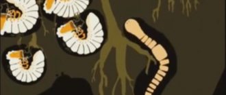
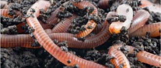
Belt of earthworms
An important role in the exchange of germ cells is played by the girdle - a small thickening on the body of the worm. When mating, the girdles should be opposite each other, and abundant and thick mucus is secreted, covering the bodies of the worms in the form of a muff.
A visual diagram of earthworm reproduction.
Seminal fluid with reproductive cells exits through small holes into the coupling, and the partners exchange its contents. The worms crawl in different directions, and the fluid with the germ cells remains for several days in the cells of the girdle.
Earthworms are purposefully bred on many farms.
During this time, the seminal fluid matures, the girdle again secretes mucus in the form of a cocoon, the worm removes it from its body, crawls out of it and leaves it in the ground, and the eggs fall into the mucus.
Fertilization is completed, eggs remain in the cocoon, from which small earthworms develop, absolutely similar to adults. Egg cocoons are formed once a week. They are similar in shape to lemons, measuring only 2-4 millimeters.
Juvenile earthworms are very small in size.
The color of the cocoons is usually light yellow, and when the eggs mature, they turn brown. The number of eggs laid by an earthworm in a cocoon ranges from 2 to 20; they develop in about 20 days.
Young worms are thin, like threads, their length is only 4-6 millimeters, but they grow quickly and feed on their own. Earthworms are able to lay cocoons from spring until early summer, then in autumn until November, until the soil freezes. Over the summer, fertile worms can lay 18-24 cocoons, each containing up to 24 eggs. After 7-12 weeks, young people are able to reproduce and produce equally fertile offspring. Mature earthworms weigh about 1 gram, and as they mature, they reach a weight of 10 grams.
Excretory system of flatworms
For the first time, special excretory organs (protonephridia) , which regulate osmotic pressure and remove excess water and dissolved metabolic products from the body. The excretory system is represented by branched tubules, which begin in the parenchyma with special stellate cells with bundles of cilia (such cells are called “flame”). Metabolic products are absorbed by the surface of the cells, and the cilia conduct fluid into their processes - excretory tubules, which combine into large ducts. Excretion products are released into the external environment through special pores.
What does cyclicality depend on?
The reproduction of worms is influenced by a lot of factors, here are the most common of them:
- Massive varieties mature for reproduction later than small ones. For example, in Dendroben, sexual maturity occurs on average from the age of 100-130 days, in Red Californians - 90-100, and Prospectors reproduce already at 80-90 days.
- The cycle is also influenced by the individual characteristics of the body. Some worms form a belt as early as 1.5-2 months, while others take up to 150-160 days.
- Ambient temperature is the first factor affecting the rate of development of cocoons and worms. The favorable temperature is +15-+25°C, when reproduction continues continuously. At low temperatures, the cocoons lie in the substrate for many months, and the worms are in suspended animation, which means they do not feed or grow.
- Availability of food. Without proper nutrition, the development of worms is delayed.
- Favorable, stable environment, without sudden changes in temperature, acidity and humidity. Stress inhibits the vital activity of worms and often leads to the death of livestock.
- Having a partner. There is an opinion that in nature worms reproduce less often than in a composter, because they do not always find a mate in time.
In captivity, the cyclical nature of reproduction is higher, as breeders strive to speed up the process, so they artificially maintain an acceptable environment. If you breed wild dung beetles, rather than selective worms, in a composter, then in 1-2 years they will become domesticated and begin to reproduce on a par with hybrids.