Fish or not
To determine the place of today's heroine in the animal kingdom, let's turn to Wikipedia. From the materials of the corresponding article we learn that lampreys belong to the class of lampreys and fish, if they are related, they are very distant, despite some external similarity.
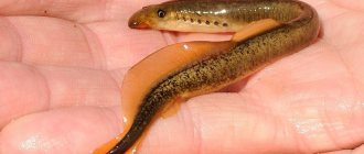
Lamprey in appearance resembles fish such as eel or loach.
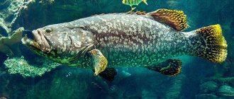
If you look closely at the photo above, you can see seven holes behind the eyes. They serve the animal for breathing, since under them there are peculiar gills.
Interesting! For these holes, the fish received a second name - seven-hole!
Features of character and lifestyle
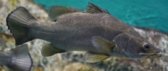
Photo: Sea lamprey
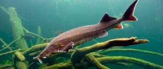
Although the lamprey fish is a predator, it leads a sedentary, lazy lifestyle. Basically, the lamprey lies at the bottom of the water pool and waits for a possible victim to swim by, to which the lamprey can attach itself. If there are no fish in the area for a long time, and the lamprey feels hungry, then it may begin to move in search of food.
Several cases of lamprey attacks on humans have been recorded. Neither was overly traumatic for people, but in both cases the victims went to hospitals for help.
Lampreys often feed on the leftovers of other fish, essentially being scavengers. They willingly eat dead tissue that falls to the bottom. Lampreys rarely swim from place to place, although they are capable of covering long distances on their own, which requires them to spend a lot of energy. Most often, lampreys travel by clinging to large fish for several days - thanks to this method, they have spread throughout almost the entire world's oceans.
Lampreys are voracious, but not aggressive. Despite the fact that they do not miss any opportunity to eat, they do not defend their territorial rights and do not conflict with other lampreys and fish that are not of nutritional interest to them. If the lamprey itself becomes someone's food, it cannot fight back the attacker.
Lampreys are solitary creatures, but they are mostly found in clusters on the bottom. This may be due either to food items that several lampreys have chosen for themselves at once, or to the spawning period.
Description
The lamprey has a long, elongated body, reaching a meter in length. The top is covered with mucus; unlike fish, there are no scales. Skin color depends on where the animal lives:
- the river dweller has gray or dark blue skin;
- marine inhabitants are most often bronze-bodied.
The animal's belly varies in color from white to dirty gray.
The structure of the animal’s oral apparatus is interesting. It is a funnel bordered by annular cartilage.
Functionally, the lamprey's mouth is a rather powerful suction cup.
Thanks to this structure of the mouth, the lamprey is able to attach itself to fish or aquatic animals. At the same time, small teeth clean off the skin of the victim and get to the meat. Often an animal affected by a lamprey dies.
On a note! Since the mouth is not involved in breathing, the lamprey can stay on someone else’s body for a long time.
Lamprey - what is it?
In our case, the Siberian lamprey ( Lethenteron kessleri
(Anikin)) is a representative of the genus of Pacific lampreys of the lamprey family. Belongs to the non-parasitic jawless chordates. Most representatives of the type have already become extinct by our time. There are only 39 species of lampreys left on the list.
In appearance, it is impossible to confuse the lamprey with another representative of the river animals.
Photo of a Siberian lamprey taken by SM Chuprov (Siberian Federal University)
When you first meet it, what catches your eye is its elongated, slippery, dense body, slightly thickened front part with 7 breathing holes. For this number of spiracles, the lamprey is popularly nicknamed “seven-hole.”
Quite large eyes are found in adults and older larvae. Has no jaws. The mouth is represented by a suction funnel. The tongue and the surface of the “mouth” are covered with small horny teeth.
It grows to a maximum length of 25 cm.
Habitat
A description of a fish will be incomplete if you do not indicate where it lives. Depending on the species, the lamprey lives in both fresh and salt water. At sea, the animal is located near the mouths of flowing rivers; movement into the open sea does occur, but not often.
The animal is found in the basins of such seas as:
- Northern;
- Baltic;
- Mediterranean.
Therefore, fish can be caught in Russia, Belarus, France, Italy, Great Britain and many other European countries.
In a reservoir, the lamprey sticks to the bottom or swims, clinging to some fish. The larvae of the animal, which are called sandworms, live buried in bottom sediments or thickets of aquatic plants and macrophytes.
Two lampreys attached themselves to this fish at once; without human help, death is inevitable.
On a note! Scientists believe that suction to fish is used by lampreys not only for feeding, but also for moving over long distances.
Distribution and habitats
The river form of lamprey is common in river basins flowing into the Baltic and North Seas. Lampreys are found in Karelia, Finland, Sweden and England. In Russia, this fish is found in Lakes Onega and Ladoga, in the rivers Luga, Narva and Neva, Voronezh. Individuals are found at depths of up to 100 meters.
Marine forms are found mainly in coastal waters of the seas. They are found from Gibraltar to Iceland, as well as all the way to the White Sea. There are populations in the Adriatic and Mediterranean Sea, off the coast of North America. There are no lampreys in the Black Sea. Sandfish, as the fish larvae are called, live in rivers that flow into the sea for several years, and only then do they swim away to live in the sea.
Nutrition
What does kaluga fish look like (photo)
Despite its parasitic nature, the lamprey feeds mainly on carrion and other organic matter found in the silt. It is easier for an animal to attach itself when there is a school of fish nearby, for example, cod, herring or mackerel. Small species even choose smelt as prey.
Being at the larval stage, the lamprey has in its diet:
- seaweed;
- worms;
- crustaceans;
- shellfish
It should be noted that lampreys themselves are food for other underwater inhabitants. She is not averse to feasting on such bottom-dwelling predatory fish species as:
- burbot:
- catfish;
- acne.
Natural enemies and species status
Lamprey is a fairly large predator. However, she also has enemies. The seven-hole fish serves as a food source for larger species of fish and crustaceans. Of the numerous larvae, only a small part of them survive to adulthood, since they are often eaten by other inhabitants of the reservoir.
The fish that the lamprey feeds on can also be enemies. She can become food for the salmon she feasted on herself.
Lampreys do not have good relationships with all feathered representatives of the fauna. In shallow areas, herons and storks often retrieve seven-hole fish from under the silt. This happens in the daytime, when she buries herself in the silt, hiding from the sunlight that irritates her eyes. They can also be caught by diving birds such as cormorants.
Lampreys often become prey for burbots that live at the bottom of reservoirs. In sea waters during the winter season, belugas and other large fish pose a danger to it. Sometimes the semidyr becomes prey for Caspian seals and other mammals that live in water bodies.
Over the past decades, there has been a sharp decline in the lamprey population, caused by the construction of hydraulic structures and river pollution. In addition, the number of fish on which lampreys parasitize has decreased significantly.
Spawning
Lampreys spawn at 3-5 years of age, depending on the species. At the same time, they stop feeding, the entire belly is left for growing eggs and milt, as a result of which the esophagus and intestines are completely degraded.
The spawning takes place on the rapids of rivers in such a way that the eggs and fry are carried by the current to their usual habitats. The process occurs on dark nights due to the fact that animals have increased light sensitivity and can interrupt the reproduction process even during the full moon or adjacent phases of the moon.
At the beginning of spawning, males clear the area at the bottom of grass, stones and other debris. They do this work with the help of the same suction cups, pulling the interference away from the nest. Females watch the process, barely touching their men’s bodies.
After this, the spawning process occurs, clearly shown in the next photo.
During spawning, females attach themselves to the selected stone, and males wrap their bodies around it, squeezing out and fertilizing the eggs.
Attention! The number of eggs of the river lamprey ranges from 14 to 40 thousand pieces. They are approximately one millimeter in diameter.
After spawning, the parents die, having completed their destined development cycle. The fry appear after a couple of weeks; depending on the water temperature, the timing shifts in one direction or another.
The larvae reach three millimeters in size and live mainly in bottom silt and sand. Even some scientists distinguish them into a separate species called sand sanders.
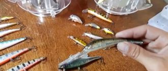
Sander is an excellent bait for catching predatory fish.
3-5 days after birth, the larvae grow approximately twice as large and burrow vertically into the sand, literally screwing into it. As a result, the slightly grown sandmoths descend downstream to the usual habitats of the species.
Kartashev N.N., Sokolov V.E., Shilov I.A. Workshop on vertebrate zoology M., Higher School. 1981
SUBTYPE VERTEBRATES - VERTEBRATA SECTION Jawless - AGNATHA CLASS cyclostomes - CYCLOSTOMATA TOPIC 2. STRUCTURE OF THE RIVER LAMPREY SYSTEMATIC POSITION OF THE OBJECT Subtype
Vertebrates, Vertebrata Class Cyclostomes, Cyclostomata Subclass Lampreys, Petromyzones Representative - River lamprey, Lamp etra fluviatilis L.
MATERIAL AND EQUIPMENT
For one or two students you will need: 1. Lamprey cut in the dorsoventral direction along the midline and fixed in alcohol. 2. Cross section of a lamprey in the region of the gill pouches. 3. Cross section of a lamprey in the intestinal area. 4. Bath. 5. Preparation needles - 2. 6. Hand magnifying glass 4-6x.
EXERCISE
Examine the appearance of the lamprey and study the structural features of the internal organs, first in longitudinal and then in transverse sections of its body. Make the following drawings: 1. Side view of the lamprey. 2. Location of internal organs on a longitudinal section of a lamprey. 3. Cross section of a lamprey in the region of the gill pouches. 4. Cross section of a lamprey in the intestinal region.
Rice. 6. Appearance of the river lamprey: 1 - oral (suction) funnel, 2 - unpaired nostril, 3 - eye, 4 - external openings of the gill sacs, 5 - lateral line organs, 6 - anus, 7 - urogenital papilla, 8 - dorsal fins, 9 - caudal fin, 10 - myomer, 11 - myosepta APPEARANCE
The river lamprey has an elongated serpentine body, divided into a head, body and tail without clear boundaries. The head and body are round in cross-section, and the tail is laterally compressed.
In front there is a suction oral funnel (Fig. 6.1), surrounded by skin papillae and having horny “teeth” on the inner surface (Fig. 7). The location and number of horny “teeth” are of systematic importance.
Rice. 7. Oral funnel of the river lamprey: 1 - oral opening, 2 - end of the tongue, 3 - horny dental plate at the end of the tongue, 4 - upper (supraoral) horny dental plate, 5 - lower (suboral) horny dental plate, 6 - upper labial "teeth", 7 - lateral labial "teeth", 8 - small marginal labial "teeth", 9 - leathery fringe of the edges of the funnel
In the depths of the funnel there is an oral opening (Fig. 7, 1) and the end of the tongue is visible (Fig. 7, 2), armed with a complex horny dental plate (Fig. 7, 3). On the upper surface of the head there is an unpaired nostril (Fig. 6, 2) and behind it an area of unpigmented skin in the area where the parietal organ is located, which serves as an additional light-sensitive organ. Paired eyes (Fig. 6, 3), located on the sides of the head, are covered with translucent skin. Behind the eyes, 7 pairs of external openings of the gill sacs open (Fig. 6, 4). The lateral line organs are located in the skin of the head and torso (Fig. 6, 5); they are noticeable in the form of mucus tubercles covering the external openings of the channels located in the thickness of the skin. On the ventral surface of the body, at the border of the body and tail, there is an anal opening (Fig. 6, 6), and behind it there is a small urogenital papilla (Fig. 6, 7).
On the dorsal surface of the body there are two dorsal fins (Fig. 6, the remains of an unpaired fin fold. The posterior end of the body is bordered by the caudal fin (Fig. 6, 9). The axial skeleton divides the caudal fin into two equal parts. Such a primary equilobed fin belongs to the protocercal type.
The bare skin of the lamprey is covered with mucus, abundantly secreted by special skin glands. The skin does not contain scales or any other elements of the exoskeleton.
The muscles of the trunk and tail consist of curved segments of myomeres (Fig. 6, 10), separated by myosepta (Fig. 6, 11). Myomeres become clearly visible if the skin is removed from a small area of the lamprey’s body.
STRUCTURE OF INTERNAL ORGANS
Skeleton. Formed by cartilage and connective tissue. The functions of the axial skeleton are performed by a dense chord (Fig. 8, 1; Fig. 9, 1; Fig. 10, 13), which has a rounded cross-section; it is surrounded by a thick connective tissue membrane (Fig. 8, 2; Fig. 9, 2). Up from the lateral sections of the notochord there are small paired cartilages - the rudiments of the superior neural arches (Fig. 9, 3; Fig. 10, 14), which laterally limit the cavity in which the spinal cord is located.
Rice. 8. Longitudinal section of a river lamprey: 1 - notochord, 2 - connective tissue membrane of the notochord, 3 - brain skull, 4 - cartilages of the oral funnel, 5 - pericardial cartilage, 6 - myomere, 7 - myosepta, 8 - musculature of the tongue, 9 - brain , 10 - spinal cord, 11 - olfactory sac, 12 - pituitary outgrowth, 13 - oral cavity, 14 - esophagus, 15 - intestine, 16 - anus, 17 - liver, 18 - gill sacs, 19 - respiratory tube, 19a - internal openings of the gill sacs, 20 - atrium, 21 - ventricle, 22 - venous sinus, 23 - abdominal aorta, 24 - kidney, 25 - ureter, 26 - urogenital sinus, 27 - gonad, 28 - urogenital papilla, 29 - genital pore
The lamprey skull, like all vertebrates, is divided into two sections: the axial, or cerebral, skull (neurocranium) and the visceral (splanchnocranium).
The cranium is the skeletal structure that protects the brain and sensory organs. In the lamprey, it is represented by a trough-shaped cartilaginous formation that protects the brain from below and from the sides (Fig. 8, 3; Fig. 10, 1); the occipital part does not develop. The olfactory capsule is adjacent to the skull in front (Fig. 10, 2), and the auditory capsules are adjacent to the sides (Fig. 10, 3). The lateral walls of the brain skull form weakly defined depressions - orbits, bounded below by the infraorbital arch (Fig. 10, 4).
Rice. 9. Cross section of a river lamprey A - in the region of the gill sacs; B - in the intestinal region: 1 - notochord, 2 - connective tissue membrane of the notochord, 3 - cartilaginous rudiments of the upper arches, 4 - myomere, 5 - myosepta, 6 - spinal cord, 7 - dorsal aorta, 8 - tongue cartilage, 9 - tongue musculature , 10 - esophagus, 11 - gill sac, 12 - external opening of the gill sac, 13 - internal opening of the gill sac, 14 - respiratory tube, 15 - abdominal aorta, /6 - intestine, 17 - spiral valve, 18 - kidney, 18a - ureter, 19 — gonad, 20 — fin ray, 21 — lymphatic cavities, 22 — posterior cardinal veins Fig. 10. Lamprey skeleton A - top view; B - side view. Brain skull: 1 - cranium, 2 - olfactory capsule, 3 - auditory capsule, 4 - infraorbital arch. Visceral skull: 5 - annular cartilage, 6 - anterior superior cartilage, 7 - posterior superior cartilage, 8 - lateral cartilages, 9 - rod-shaped cartilages, 10 - hyoid cartilage, 11 - gill lattice, 12 - pericardial cartilage. Axial skeleton: 13 - notochord, 14 - rudiments of neural arches
The visceral skull is the skeletal formation that develops in the walls of the anterior part of the digestive tube. Functionally, these formations represent the skeleton of the oral and gill apparatus. In the lamprey, the visceral skull includes cartilages that support the oral funnel (Fig. 8, 4; Fig. 10, 5-9) and the tongue (Fig. 10, 10), as well as an openwork cartilaginous lattice that supports the region of the gill sacs (its the structure is clearly visible on specially prepared skeletons, Fig. 10, 11). In the posterior part of the visceral skull there is pericardial cartilage (Fig. 8, 5; Fig. 10, 12), surrounding the heart from behind and on the sides and connecting to the peribranchial lattice by cartilaginous bridges.
The fins of the lamprey are supported by thin cartilaginous fin rays (Fig. 9, 20). Musculature. The lateral muscles of the trunk and tail are formed by muscle segments - myomeres (Fig. 8, 6), which have a complex shape and are separated from each other by myosepta (Fig. 8, 7; Fig. 9, 5). In the gill region, the muscular cords bifurcate, forming the epibranchial and subbranchial muscles. In the head region of the lamprey, the complexly differentiated muscles of the tongue are well developed (Fig. 8, 8; Fig. 9, 9). The central nervous system is clearly divided into the brain (Fig. 8, 9) and the spinal cord (Fig. 8, 10; Fig. 9, 6). The spinal cord has a ribbon-like flattened shape, is located above the notochord and is covered from above and from the sides by the connective tissue membrane of the notochord.
The external unpaired nostril communicates with a round, dark-colored, folded olfactory sac through a short nasal canal (Fig. 8. 11). A blind pituitary process extends from the olfactory sac down and back under the brain and the anterior part of the notochord (Fig. 8, 12). The pituitary outgrowth, together with the nasal canal, serves as a kind of pipette that sucks and expels water from the olfactory sac. Digestive system. The oral opening, located in the depths of the suction funnel, leads into the oral cavity (Fig. 8, 13). The lamprey has a pharynx only at the larval stage. During metamorphosis, it is divided into two independent sections: the esophagus and the respiratory tube (see description of the respiratory system). The esophagus (Fig. 8, 14; Fig. 9, 10) starts from the back of the oral cavity, goes under the notochord and, going around the heart, imperceptibly passes into the intestine (Fig. 8, 15; Fig. 9, 16). The intestine stretches over the liver, then goes back without bends along the lower surface of the abdominal cavity and ends at the anus (anus). The inner part of the intestinal mucosa forms a longitudinal fold protruding into the intestinal cavity - it is called a spiral valve (Fig. 9, 17); it increases the absorption surface of the intestine.
Behind the heart is a large compact liver (Fig. 8, 17). Adult sea-dwelling lampreys have a gall bladder and bile duct; in individuals that go to rivers to spawn and stop feeding, they are reduced. Lampreys, when feeding, stick to the body of the prey (fish) with their mouth funnel. Horny “teeth” located on the inner surface of the oral funnel contribute to a more durable attachment of the lamprey. Using a horny dental plate at the tip of the tongue, the lamprey makes a hole in the skin of the victim. Thanks to the rhythmic contractions of the powerful muscular tongue, which acts like a piston, blood is sucked into the oral cavity and from there enters the esophagus. Respiratory system. Unlike all other vertebrates, cyclostomes develop gill sacs in the gill slits (Fig. 8, 18; Fig. 9, 11), which are of endodermal origin. The inner surface of the gill sacs forms numerous folds of the mucous membrane, in which there is a dense network of small blood vessels - capillaries. In the lamprey, each gill sac (there are 7 pairs in total) opens outward with an independent external opening (Fig. 6, 4; Fig. 9, 12). The internal opening of the gill sac (Fig. 9, 13) connects its cavity with the respiratory tube (Fig. 8, 19; Fig. 9, 14), which is a blind outgrowth connected in front to the oral cavity and delimited from it by a movable fold - a sail .
In a swimming lamprey, water enters the oral cavity, from there it passes into the respiratory tube and, having passed through the gill sacs, is thrown out through their external openings. In this case, oxygen dissolved in water penetrates the capillaries and is bound by blood pigment, and carbon dioxide saturating the venous blood passes into the water and is removed with it.
In the case when the lamprey is feeding (or if it has attached itself to any object), the flow of water through the oral cavity becomes impossible. At the same time, the sail closes the entrance to the breathing tube, preventing water from passing through, and liquid food passes from the oral cavity into the esophagus. In this case, breathing is carried out in a different way: under the influence of the muscles of the body walls, the gill sacs are compressed, and water is pushed out through the external gill openings (active exhalation); further, thanks to the elasticity of the cartilaginous gill lattice, the gill region expands again, and water through the same external openings is again sucked into the gill sacs (passive inhalation). Circulatory system. Cyclostomes have a well-developed heart, the contractions of which ensure blood circulation throughout the body. The heart (cor) is located behind the last pair of gill sacs and is delimited from the underlying liver by the pericardial cartilage (Fig. 8, 5). The lamprey's heart is two-chambered: it consists of one atrium (atrium; Fig. 8, 20; Fig. 11, 2) and one ventricle (ventriculus; Fig. 8, 21; 11, 3), and behind them is the venous sinus (sinus venosus ; Fig. 8, 22). Venous blood enters the venous sinus through the veins, from there it passes into the atrium and then into the ventricle. A powerful arterial stem departs from the ventricle - the abdominal aorta (aorta ventralis; Fig. 8, 23; Fig. 11, 5), which branches into afferent gill arteries, through which venous blood enters the capillaries of the folds of the gill sacs. Oxidized (arterial) blood is collected through the efferent gill arteries into the unpaired dorsal aorta (aorta dorsalis; Fig. 9, 7; Fig. 11, 6) and is distributed throughout the body along its branches.
Fig. 11. Scheme of the circulatory system of the river lamprey (ventral view): 1 - venous sinus, 2 - atrium, 3 - ventricle, 4 - aortic bulb, 5 - abdominal aorta with afferent branchial arteries extending from it, 6 - dorsal aorta, 7 - efferent branchial arteries flowing into the dorsal aorta, 8 - anterior cardinal vein, 9 - inferior jugular vein, 10 - tail vein, 11 - posterior cardinal vein, 12 - intestinal vein, 13 - portal system of the liver, 14 - hepatic vein
From the anterior part of the body, venous blood is collected by the anterior cardinal veins, and from the powerful muscles of the tongue - by the inferior jugular vein (Fig. 11). The tail vein in the body cavity is divided into paired posterior cardinal veins. The subintestinal vein collects blood from the intestines; it then enters the liver and breaks up into capillaries (portal system of the liver), and the hepatic capillaries merge into the hepatic vein. All these large veins (anterior and posterior cardinal, inferior jugular, hepatic) flow into the venous sinus. Thus, cyclostomes, like skullless ones, have one circle of blood circulation. The complete circulatory system can only be seen with special injected preparations; on the cross sections considered, only individual vessels are visible (Fig. 8 and 9). Genitourinary system. The excretory organs of cyclostomes are the mesonephric (trunk) kidneys (ren; Fig. 8, 24; 9, 18), which in the form of paired ribbon-like formations are suspended on the mesentery to the dorsal wall of the posterior half of the body cavity. A thin canal, the ureter, runs along the lower edge of the kidneys. At the posterior edge of the kidneys, the ureters of the right and left sides connect and form an ampulla-shaped extension - the urogenital sinus (Fig. 8, 26), which opens outward at the apex of the urogenital papilla (Fig. 8, 28).
The gonad (Fig. 8, 27; Fig. 9, 19) is unpaired and occupies almost the entire free part of the body cavity. The ovaries of females are clearly distinguishable from the testes of males by their granular structure. There are no reproductive ducts; sexual products enter the body cavity through ruptures in the wall of the gland, then through the genital pores (openings in the walls of the urogenital sinus) flow into the urogenital sinus and are discharged out through its opening.
CONCLUSION
Cyclostomes - the first class of the subphylum of vertebrates (Vertebrata) - have a number of progressive structural features compared to lower chordates (including skullless ones). Differentiation of the anterior part of the neural tube into the brain with several morphologically and functionally separate sections leads to improved nervous control over various functions of the body, which provides the possibility of more active life. The separation of the brain and sensory organs requires the emergence of protective formations that protect these important organs from mechanical damage. This is ensured by the formation of a cartilaginous skull, which was absent in lower chordates. The rudiments of the upper vertebral arches developing above the notochord perform the same function in relation to the spinal cord. The appearance of the visceral skull in the form of cartilages supporting the oral funnel and pharynx facilitates the transition to active nutrition and intensifies breathing. The respiratory function is also significantly intensified by the appearance of specialized respiratory organs in the form of gill sacs, which increase the possibility of active respiratory movements.
Numerous folds on the inner surface of the gill sacs, which has a dense network of capillaries, greatly increase the total respiratory surface, and therefore the amount of oxygen entering the blood.
The intensification of gas exchange both in the gills and in the tissues is also facilitated by the formation of the central circulatory organ - the heart, the contractions of which lead to faster blood flow through the vessels. Finally, a new type of excretory organs, the kidneys, appears, which distinguishes cyclostomes (as well as other vertebrates) from lower chordates. Mesonephric (trunk) kidneys, functioning in adult cyclostomes, cope more efficiently with the function of excretion at an increased level of metabolism than, for example, the nephridial system of the lancelet. In the kidneys, dissimilation products are filtered not only from the cavity fluid, but also from the blood. The listed structural features indicate that cyclostomes retain the structure plan that arose in the subtype of skullless, but as a result of complication and differentiation practically. of all organ systems, they have developed a higher level of organization, providing a more active lifestyle and a higher level of metabolism. All these features: the development of the brain, skull (axial and visceral), spine (even in its most rudimentary form), heart, kidneys, specialized respiratory organs - are characteristic of all vertebrate animals and represent characteristic morphological features of this subtype. Compared to other classes of the vertebrate subphylum, cyclostomes have many primitive features that significantly distinguish this group from the other six classes. Such signs include the absence of paired limbs, preservation of the notochord throughout life and the absence of formed vertebrae, a simple structure of the brain skull, still open at the top and incompletely fused with the capsules of the sensory organs, the absence of the genital ducts and some others. The specific features of cyclostomes are especially noticeable in the structure of the visceral skull and respiratory organs. Of all vertebrate animals, only cyclostomes have gills of endodermal origin and have the form of bags. In fish (see p. 44), the gills are ectodermic and are formed by many gill filaments, attached to the elements of the visceral skeleton (gill arches) and with the free end hanging into the cavity of the gill slit, through which the water flows. The visceral skeleton of cyclostomes is represented by cartilaginous formations that have a supporting function and do not form dissected movable arches, as in other vertebrates. Accordingly, the skeleton of the oral apparatus of cyclostomes does not have movable jaws, and the skeleton of the gill region does not have movable gill arches. The sum of all these characteristics required the division of cyclostomes (including fossil forms) into a special section of the vertebrate subtype, which, due to the absence of jaws, was called Agnatha (jawless), as opposed to the section Gnathostomata (gnathostomes), which includes the remaining classes. It is easy to see that gnathostomes have great potential for activating their lifestyle and intensifying metabolism: their visceral skeleton, in addition to performing supporting and protective functions, takes a much greater part in active acts of feeding (grasping and other movements of the jaws) and breathing (movements of the gill arches, which contribute to more active breathing). Apparently, this circumstance was the reason that in the evolution of vertebrates, the group of gnathostomes quite quickly replaced the once numerous jawless animals. Currently living cyclostomes have occupied a special ecological niche (especially in terms of the type of nutrition), which has given them the opportunity to get out of competition with gnathostomes even with relatively inactive life activity. In turn, the environmental characteristics of these animals affected their structure. It has already been said above that the division of the pharynx into the pharynx itself and the respiratory tube in lampreys is associated with the peculiarities of their nutrition. The same is the adaptive significance of the sail, horny “teeth” on the inner surface of the suction funnel, a powerful muscular tongue, etc. In hagfish, which, when feeding, “bite deeply” into the body of the prey, the outer gill openings are covered with paired skin folds, due to which the common gills the holes are “pushed back” far back. This allows the hagfish to breathe during the act of feeding, which lasts for a long time. The structural features of modern cyclostomes reflect the general progressive evolution of chordates, as well as the adaptation of modern forms to specific conditions of existence and way of life. This pattern is seen in other vertebrate groups.
additional literature
Balabay P.P., Morphology and phylogenetic development of the group of agnathans, Kyiv, 1956. Gurtovoy N.N., Matveev B.S., Dzerzhinsky F.Ya. Practical zootomy of vertebrates. Lower chordates, jawless fish. M., 1976. Shmalgauzen I.I. Fundamentals of comparative anatomy of vertebrates. M., 1947.
BackContentsNext
The nutritional value
The nutritional value of lamprey is high, but the animal is also valued for its unusual taste, appreciated by gourmets. We present the characteristics of its meat in the table.
| Characteristic | Quantity per 100 grams |
| Calorie content | 88 kcal |
| Squirrels | 18 g |
| Fats | 2 g |
| Carbohydrates | 0 |
Lamprey is fried, stewed, baked, boiled, pickled.
Danger to humans
Lampreys very rarely attach themselves to humans, so you should not be especially afraid of them. However, their bite is quite unpleasant due to special substances in the animal’s saliva that prevent blood clotting. Doctors advise in such cases to immediately contact a medical facility to avoid the possible destruction of red blood cells and prevent tissue breakdown.
And it also needs to be said about the toxicity of the mucus covering the animal’s body. To avoid poisoning, you should carefully remove it before cooking, or better yet, remove the skin altogether.
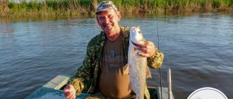
Advice! To avoid painful troubles, you need to hold a live lamprey carefully, as the person in the next photo does.
You need to hold it, making sure to grab the place behind the animal’s head with your hand.
Fishing methods
Lampreys don't take the bait! Fishing only with various traps:
- networks;
- muzzles;
- tops;
- lionfish.
The main fishing time is during the spawning run, when it is most productive in terms of financial and human costs.
Taking advantage of the fact that the animal is afraid of light, they practice this method of catching:
- A section of the river in a deep place is blocked with an extended net.
- At night, lights begin to shine to the right and left of the set trap.
- Lampreys, fleeing from the rays, gather towards the unlit center and, like a tunnel, end up in a net.
Fishing
Ukrainian river lamprey is quite rare, so its use in fishing is virtually impossible. The fish itself is not of interest to fishermen in its pure form.
However, it is very often used as bait for larger and more predatory fish. It is not difficult to catch this type of lamprey; you just need to take into account the characteristics of the character and behavior of the species.
Given the fact that adults do not feed, you can catch them using small nets or using algae-based bait to attract young individuals that are still in the larval stage.
Fishing for lampreys should be done on a sandy-clay bottom with a fast, cold current. In such places the greatest concentration is observed.